The Nanobiotechnology Laboratory is a shared laboratory at the CEITEC Instruct centre helping other scientists, such as biochemists, chemists and biologists, to better understand the biological processes taking place in living cells, but also to image biomolecules in their natural environment, or complex properties of new nanomaterials. Our labs contribute to a better understanding of heart diseases, the effects of cytostatics on cancer cells or to the development of new treatments of osteoarthritis. We monitor the structure of biomolecules and nanomaterials for the preparation of new types of biosensors. Monitoring cell adhesion then contributes to the development of new implant materials. The main instruments used in our laboratories are AFM microscopes, confocal optical microscope, Raman microscope, biosensor platforms, electrochemical and electrophysiological instruments.
More information can be found on the Nanobiotechnology core facility (NANOBIO) webpage.
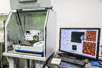
JPK NanoWizard® 4XP combined with a fluorescence microscope (incl. FluidFM module and Bruker BioSoft NanoIndenter).
JPK NanoWizard® 4XP AFM combined with a fluorescence microscope Leica DMi 8 (incl. FluidFM module and Bruker BioSoft NanoIndenter). Extended range of operation (200 x 200 x 200 um) offers unique possibility to characterize structural and mechanical properties of large samples, such as tissue sections. Leica DMi 8 inverted optical microscope is used for the most of measurements, however, the top-view optics gives possibility to work with non-transparent samples, too.
AFM modes: contact mode, AC mode (tapping), quantitative imaging (QI), Force Mapping, etc.
FluidFM module in combination with AFM features can be used as nano-pipette – to either inject or to aspirate nanolitre volumes, or to test cell adhesivity. Bruker Biosoft indenter can be used for standard indentation studies, now on a very soft sample (cells, tissues, hydrogels).
More information about the instrument available here.
The Dimension FastScan Bio Atomic Force Microscope (AFM) enables high-resolution research of biological dynamics, with temporal resolution of up to 3 frames-per-second for live sample observations. What’s more, it does this while making the AFM easier to use than ever before. Built upon the world’s most advanced large-sample AFM platform, the FastScan Bio AFM adds specialized life science features to this platform for high-resolution, live-sample observation of interacting molecules, membrane proteins, DNA protein binding, inter-cellular signaling and many other dynamic biological studies.
AFM modes: contact mode, standard tapping mode, Peak-Force Quantitative Nanomechanics (PFQNM) and Force Volume.
More information about the instrument is available here.
The MultiMode® platform's long history of success is based on superior resolution, performance, and unparalleled versatility and productivity. The MultiMode 8-HR atomic force microscope (AFM) further advances these capabilities to improve imaging speed, resolution significantly, and nanomechanical performance with higher speed PeakForce Tapping®, enhanced PeakForce QNM®, new FASTForce Volume, and exclusive Bruker probes technology.
More information about the instrument is available here.
JPK NanoWizard® 3 combined with a fluorescence microscope and Olympus FV2100 confocal microscope.
The proven concept of the bioAFM microscope from JPK, serves well for combined measurements with the optical microscope from Olympus. Thus, combined measurements can be performed where the AFM provides information on the mechanical properties of the sample and the fluorescence microscope simultaneously displays the structural details.
AFM modes : contact mode, AC mode (tapping), quantitative imaging (QI), Force Mapping, etc.
More information about the instrument is available here.
NTMDT Solver Nano is equipped with a professional 100 micron CL (closed loop XYZ) piezotube scanner with low noise capacitance sensors. Capacitance sensors in comparison with strain gauge and optical sensors have lower noise and higher speed in the feedback signal. The CL scanner is controlled by a professional workstation and software.
More information about the instrument is available here.
The MEA2100-Mini-System is a miniaturized in vitro recording system. The system can be operted continuously in an incubator while maintaining most of the MEA2100 platform advantages. The system consists of a 60-channel headstage, featuring a sampling rate of 50kHz per channel at a 24bit resolution, as well as a 2-channel stimulator with user customizable patterns. The digitized data is transfered from the headstage to a signal collector unit for up to four headstages.
An easy combination of this MEA system with the AFM (JPK NW3/4) gives a unique opportunity to study mechano-electrical coupling/decoupling phenomenon of the cardiac cells contraction.
More information about the instrument is available here.
Raman microscope will give you:
Highly specific identification - Raman microscopy can differentiate chemical structures, even closely related ones. High spatial resolution - comparable to your other microscopic techniques (confocal pinhole available). Particle analysis - use advanced image recognition algorithms and instrument control features to characterise particle distributions. Correlative imaging - create composite images by combining Raman data with either optical (upright) or AFM microscope.
More information about the instrument is available here.
The instrumentation available in the CF laboratory allows structural and biomechanical studies at the level of single molecules, through single cells to tissue sections and whole organisms (e.g., young plants). For this purpose, we mainly use bioAFM instruments in combination with optical (fluorescence, confocal) microscopes. Complementary techniques are e.g., Raman microscopy for mapping chemical properties, multi-electrode arrays for characterization of electrical properties of cells or microplate readers.
The laboratory provides physical as well as remote access option. The staff supports users with planning and optimisation of the measurements.
The services are provided for the following studies:
Cells - mechanical properties
Living cells in either standard plastic Petri dish (TPP 93040) or special confocal dish (consult us for a list of compatible dishes). Force Mapping of cell cultures, mechanical characterization of cardiomyocytes. Possible combination with optical microscopy (bright field, fluorescence) – independent/overlay imaging with AFM. Standard CO2 incubator and small flow box available in the lab. UV sterilization of the room and workspace every day.
Cells - imaging
Living cells in either standard plastic Petri dish (TPP 93040) or special confocal dish (consult us for a list of compatible dishes). Fixed cells (PFA) on a glass slide. Modes: contact mode, QI, PF-QNM, Force Volume. Possible combination with optical microscopy (bright field, fluorescence) – independent/overlay imaging with AFM. Standard CO2 incubator and small flow box available in the lab. UV sterilization of the room and workspace every day.
Biomolecules - imaging
AFM imaging of biomolecules (proteins, DNA, macromolecules and biopolymers) and their complexes. Immobilization on mica (muscovite), other materials (HOPG, silicon, metal electrodes) can be used as well. Methods: tapping mode, QI, PF-QNM, Force Volume.
Nano-objects imaging
AFM imaging of nano-objects (nanoparticles, nanotubes, nanorods, etc.) and their complexes. Immobilization on mica (muscovite), other materials (HOPG, silicon, metal electrodes) can be used as well. Methods: tapping mode, QI, PF-QNM, Force Volume.
Raman-AFM combined microscopy
Raman microscopy
Mapping of chemical properties of the samples. Combined with optical and/or AFM imaging.
Electrochemical measurements
High-end electrochemical analyzer (potentiostat/galvanostat) for voltammetry, amperometry and electrochemical impedance spectroscopy (EIS) on diverse electrodes and surfaces. Possibility of 2-channel measurements, high sensitivity, low noise. SW Autolab Nova for data evaluation.
Nanodeposition system
Non-contact (ink-jet) deposition of pico- and nano- liter droplets of samples onto diverse substrates. Positioning of the droplets with micrometer precision, creation of various patterns and arrays, protein chip fabrication. Immobilization of proteins and other biomolecules on specific positions in controlled temperature and humidity.
Fluorescence scanner
Fluorescence scanning of protein, glycan, DNA, cell and other arrays or fluorescently labeled samples. The substrate dimensions are limited to microscope slides. Resolution down to 0.5 um/px. Three fluorescence channels (ex/em): 488/520, 532/572, 635/675 nm. Autosampler for 24 slides. Data evaluation in SW Mapix.
SPR biosensor
Two-channel SPR (bio)sensor based on surface plasmon resonance. Characterization of optical properties of thin layers in solution and in dry state, and measurement of their changes in real-time. The instrument is a true goniometric SPR providing a wide angular range. Use of 2 laser wavelengths allows to measure the refractive index and layer thickness. Combination with simultaneous electrochemical measurements is possible. Label-free measurement and characterization of biomolecular interactions with one binding partner immobilized on the surface, the other free in solution. Determination of kinetic parameters and binding constants, concentration measurement of various analytes.
FluidFM
Special combination of AFM with microfluidics, suitable for nanoliter injection/aspiration to/from a single cell; cell adhesion studies, single cell biomechanics/electrochemistry.
NanoIndenter studies
BioSoft indenter suitable for study of soft biomaterials and biological samples (cells, tissues, hydrogels, etc.)
The full list of instruments available:
More information is available here.

